Investigation of left ventricular myocardial deformation in patients with sarcoidosis
Sarcoidosis is a systemic granulomatous disease affecting various organs including the heart. It is characterised by the formation of granulomas, i.e. foci in the tissue in which there is an aggregation of immune cells responding to a previously unknown stimulus. Females are more likely to suffer from this disease of unknown origin than males, in a ratio of 1.5-2.0:1. Sarcoidosis of the heart is clinically quite rare, reported to affect between 2 and 7% of patients (although some sources suggest that this may be as high as 30-40%). However, it is one of the most serious forms of the disease, as it causes cardiac arrhythmias that can lead to sudden death.
Scientists at our center focused on the following in their study entitled “Left ventricular myocardial deformation assessment in asymptomatic patients with recently diagnosed sarcoidosis of the respiratory tract and/or extrapulmonary sarcoidosis“, whether myocardial deformation analysis by magnetic resonance imaging could help in the early detection of this disability.
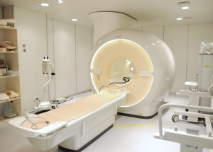
One hundred and thirteen patients with airway and/or extrapulmonary sarcoidosis without evidence of cardiovascular disease participated in the study. In these patients, myocardial strain was measured from cardiac magnetic resonance images using a technique called feature tracking (CMR-FT), followed by comparison with twenty-two healthy patients. “The main parameters measured were global longitudinal strain, global circumferential strain and global radial strain of the left ventricle,” said Assoc. MUDr. Roman Panovský Ph.D.
The results of the research conducted by scientists from the Cardiovascular Magnetic Resonance Research Team of the FNUSA-ICRC show that the values of myocardial deformation of patients with sarcoidosis did not differ significantly from the results of the control group. A possible explanation could be that the patients were investigated at an early stage of the disease when the heart muscle is not yet affected. Despite these results, the CMR-FT method has great potential for use, especially considering the fact that it offers additional information about the systolic function of the heart without prolonging the examination. The study was published in the Orphanet Journal of Rare Diseases (IF 3.7).
Supported by the European Regional Development Fund – Project ENOCH (No. CZ.02.1.01/0.0/0.0/16_019/0000868).


